Biotechnology Center Of Ho Chi Minh City


Harnessing Indigenous Bacteria toward Biological Solutions for PE, PET, and PS Plastic Waste Treatment
(SHTP) – From soil and plastic waste samples collected at several polluted sites in Ho Chi Minh City, the research team from the Environmental Biotechnology Department, Biotechnology Center of Ho Chi Minh City, isolated and selected indigenous bacterial strains capable of acting on polyethylene (PE), polyethylene terephthalate (PET), and polystyrene (PS). After 30 days of testing, several strains produced marked mass loss relative to the controls and induced changes in plastic surfaces observed by FE-SEM. The findings provide a potential microbial resource for developing biological solutions for plastic waste treatment.
From indigenous bacterial resources to the selection of promising strains
PE, PET, and PS are widely used polymers but are highly resistant to environmental degradation. The study explored microbial communities adapted to plastic waste at the Nhieu Loc–Thi Nghe Canal, Doi–Te Canal, Vam Thuat River, and Xuyen Tam Canal. Microorganisms were enriched in mineral salt medium containing PE, PET, or PS as a carbon source, and eight isolates—PE1, PE2, PET1, PET2, PS1, PS2, PS3, and PS4—were subsequently obtained. Their effects on plastics were evaluated based on mass changes after 30 days, FE-SEM imaging, extracellular enzyme activities, 16S rRNA identification, and PCR screening of selected genes associated with polymer metabolism.
Five strains—PE2, PET1, PET2, PS1, and PS2—were selected for further analysis. PE showed the clearest response: PET1, PET2, PS1, and PS2 each exhibited apparent mass losses above 50%, corresponding to net mass losses of approximately 50.31–53.31% after control correction; PE2 reached 42.23%. For PET, PS2 showed the highest net mass loss at approximately 26.46%, followed by PE2 (17.40%) and PS1 (16.16%). PS was more resistant, with PS1 showing a net mass loss of approximately 11.20%.
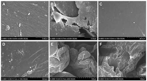
Figure 2. FE-SEM images of PS, PET, and PE surfaces after 30 days. A, C, E: abiotic controls; B, D, F: samples incubated with the microbial consortium, showing surface pits, microcracks, and delamination.
Bacterial identification and markers associated with polymer metabolism
16S rRNA analysis assigned the selected isolates to the genera Streptomyces, Cupriavidus, Pseudomonas, and Aquipseudomonas; PS1 was identified as Pseudomonas lalkuanensis (99.24%), while the remaining isolates were retained at the genus level.
From indigenous bacterial resources to the selection of promising strains
PE, PET, and PS are widely used polymers but are highly resistant to environmental degradation. The study explored microbial communities adapted to plastic waste at the Nhieu Loc–Thi Nghe Canal, Doi–Te Canal, Vam Thuat River, and Xuyen Tam Canal. Microorganisms were enriched in mineral salt medium containing PE, PET, or PS as a carbon source, and eight isolates—PE1, PE2, PET1, PET2, PS1, PS2, PS3, and PS4—were subsequently obtained. Their effects on plastics were evaluated based on mass changes after 30 days, FE-SEM imaging, extracellular enzyme activities, 16S rRNA identification, and PCR screening of selected genes associated with polymer metabolism.
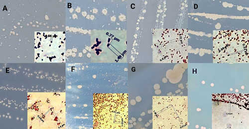
Figure 1. Colony and cell morphology of the eight bacterial isolates. Labels A–H correspond to PE1, PE2, PET1, PET2, PS1, PS2, PS3, and PS4, respectively.
Effects of Selected Bacterial Strains on PE, PET, and PSFive strains—PE2, PET1, PET2, PS1, and PS2—were selected for further analysis. PE showed the clearest response: PET1, PET2, PS1, and PS2 each exhibited apparent mass losses above 50%, corresponding to net mass losses of approximately 50.31–53.31% after control correction; PE2 reached 42.23%. For PET, PS2 showed the highest net mass loss at approximately 26.46%, followed by PE2 (17.40%) and PS1 (16.16%). PS was more resistant, with PS1 showing a net mass loss of approximately 11.20%.
Figure 2. FE-SEM images of PS, PET, and PE surfaces after 30 days. A, C, E: abiotic controls; B, D, F: samples incubated with the microbial consortium, showing surface pits, microcracks, and delamination.
Bacterial identification and markers associated with polymer metabolism
16S rRNA analysis assigned the selected isolates to the genera Streptomyces, Cupriavidus, Pseudomonas, and Aquipseudomonas; PS1 was identified as Pseudomonas lalkuanensis (99.24%), while the remaining isolates were retained at the genus level.
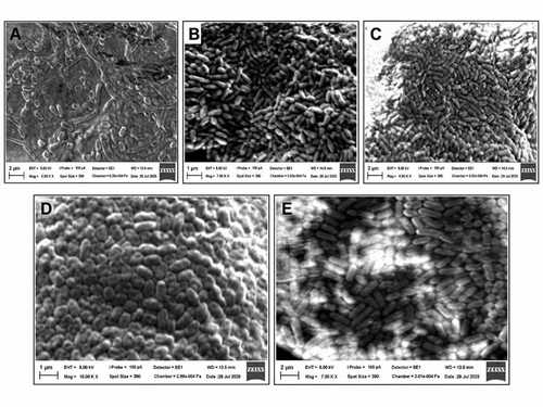
Figure 3. Cell morphology of selected bacterial strains observed by scanning electron microscopy (SEM) at 10,000× magnification. (A) Streptomyces sp. (PE2); (B) Cupriavidus sp. (PET1); (C) Cupriavidus sp. (PET2); (D) Pseudomonas lalkuanensis (PS1); and (E) Aquipseudomonas sp. (PS2). Scale bars are shown directly in each micrograph.
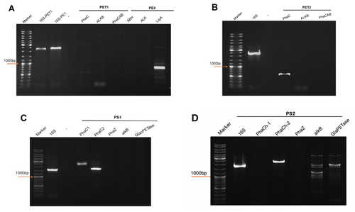
Figure 4. PCR screening of selected genes associated with polymer metabolism in the selected strains. PCR results are used as preliminary indicators and require further confirmation at the levels of gene expression, enzyme activity, and metabolic products.
Figure 4. PCR screening of selected genes associated with polymer metabolism in the selected strains. PCR results are used as preliminary indicators and require further confirmation at the levels of gene expression, enzyme activity, and metabolic products.
PCR screening detected in PS2 signals associated with PETase, alkB, and genes of the PHA cycle; PE2 showed a lipA signal, whereas the remaining strains displayed different gene combinations. These signals help guide hypotheses on metabolic mechanisms and provide a basis for subsequent confirmation of polymer-degrading functions. Further work should integrate sequencing, quantitative gene-expression analysis, enzyme activity assays, and analysis of relevant metabolic products. These preliminary findings indicate that indigenous bacteria from Ho Chi Minh City have potential for biological plastic waste treatment. The degradation mechanisms should be further validated before broader application.
Environmental Biotechnology Department
Biotechnology Center of Ho Chi Minh City
Biotechnology Center of Ho Chi Minh City
Author: Nguyen Thi Ai Niem
Source: shtp.hochiminhcity.gov.vn
Older articles
Events
-
 The 13th Asian Congress on Biotechnology (ACB 2017)...
- 26/01/2017 04:49:00 AM
-
 Welcome to the 19th A-IMBN Annual Conference 2016
- 21/04/2016 01:29:00 PM
-
 Groundbreaking Ceremony for Components of the HCMBiotech...
- 06/11/2015 10:51:00 AM
-
 Artificial Cell Demonstrates Dynamic Gene Regulation...
- 06/11/2015 10:23:00 AM
-
 Low-Cost Sophisticated Device Diagnoses HIV (10-12-2014)
- 06/11/2015 10:20:00 AM
Visitors


Photos activities


Weblinks


